Meet the scientists who walk inside cells
Nothing is more fundamental to our understanding of life and disease than the cell. We are made of them, but we still don’t know enough about them. What happens in a healthy cell? What happens to a cell in disease? What can we do to fix a cell that is unwell?
Technology is helping us to answer these questions. On the frontier of this technology is virtual reality. Virtual reality (VR) is allowing scientists to walk inside cells and observe first hand a moment frozen in time or the behaviour of a cell in real time.
Rob Parton has dedicated his career to understanding these building blocks of life and using technology to do so. He is quite simply fascinated. The first time he walked ‘inside’ a cell he didn’t want to leave.
“It’s better than I ever imagined it could be,” he said. “Cell biology has traditionally involved looking at flat images. With new 3D technology we can re–create the cell, and with VR, ‘miniaturise’ ourselves allowing us to walk inside the cell.”
Scientists are using the technology to understand better how cells work in health and disease. They take a cell that they want to study, for example, a cancer cell, place the sample inside an electron microscope and cut it into thousands of slices. As the block face is exposed, it is imaged, and these images are then put together to make a movie of the cell.
“The cell is frozen in time. We can look at all the compartments, how they interconnect and interact. We can then identify different structures in the cell that we are interested in exploring further,” said Professor Parton.
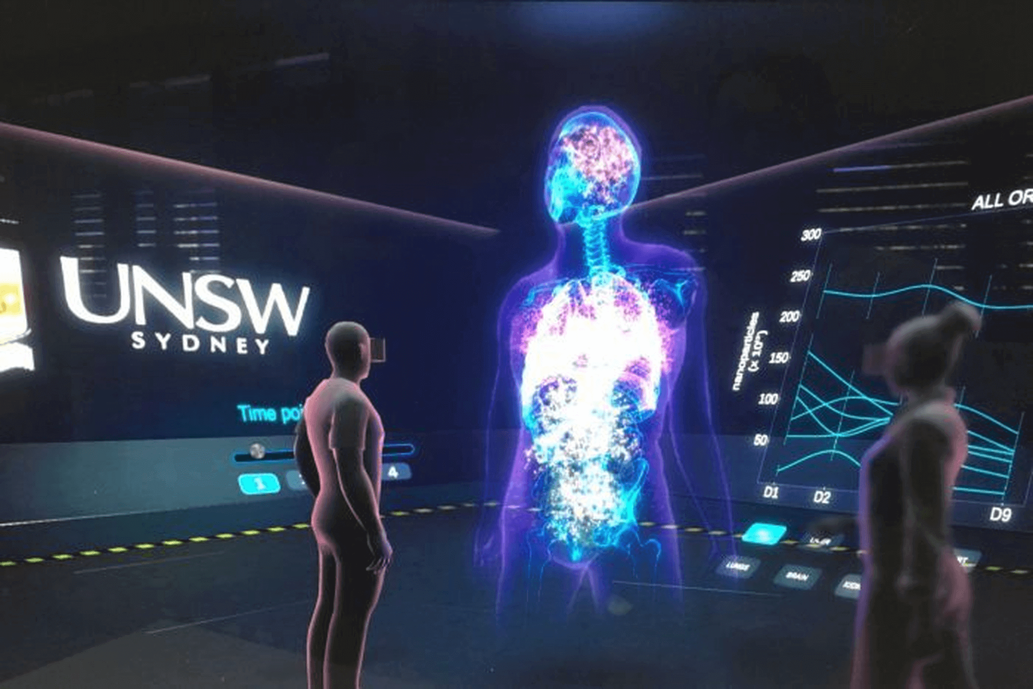
Professor Parton’s team is exploring how novel nano–particles, made in their lab, can be sent to a cell like a postage parcel. The nano–particle can enclose a drug, be addressed to a specific cell, which it then penetrates through the cell membrane to deliver the drug, to kill cancer for example. They are also exploring how they might be used to deliver vaccines.
Professor Jenny Stow is another scientist making use of this cutting edge technology to explore the cell, but she is looking at live cells – real life events, in real space and time.
“This is a revolutionary time. It is so exciting to be a cell biologist right now. Technology, particularly 4D processing, has transformed the science,” she said.
Professor Stow is using rapid, high resolution laser microscopes to image cells in 4D (3D images over time), along with super computing, to connect with VR, to visualise the intricate behaviours of molecules, cells or even small organisms. She can then see how things work (or don’t work in disease) in unprecedented detail.
“We’re looking at how bacteria and white blood cells interact during infections and at how tumour cells respond over the short and long term to drug treatments,” said Professor Stow.
“Up until now, we’ve only been able to look at these things in low resolution and in two dimensions. Now we can image live samples, reconstruct the images and through VR, study any perturbations in cell behaviour caused by drugs, infecting pathogens or gene mutations.”
Professor Stow and her colleagues utilise a new Lattice Light Sheet Microscope, invented by Nobel Prize Winner Eric Betzig.
This microscope surpasses any previous technology. It combines high–resolution imaging capacity with super fast imaging speed but does not damage the cells. It allows for extended exposure times without loss of clarity, which means that researchers can make detailed movies of processes under observation over long periods, taking their understanding to a deeper level. They will be able to watch the evolution of cell processes over several days.
“By observing cells for days, we can see them divide and move and watch the effects of disease or treatments over several cell generations. This imaging, coupled with VR, generates the comprehensive insights we need to understand the biology of cells in cancer, infection and inflammation,” said Professor Stow.
Scientists are also using new technologies to build sensors to visualise cell signals.
“Combining biosensors with breakthrough techniques like lattice–light sheet microscopy is a revolution,” said cell biologist Professor Alpha Yap.
While microscopy has principally been used to study the micro– or nano–anatomy of cells, it is now capable of studying dynamic functional processes.
“For decades scientists have been studying the signaling pathways that control how cells behave. These are often very fast (working in seconds or minutes) and we have usually studied them by using biochemical techniques, grinding up millions of cells,” said Professor Yap.
“These approaches don’t capture the fast dynamics of signaling. Nor do they tell us where in the cells signals operate. And location is important, because it is increasingly evident that when signals are mis–localized, or act on the wrong time scales, then disease can eventuate.”
Microscopes have always been central to the study of cell biology and disease. Now, these new technologies, coupled with VR, are providing Professors Parton, Yap, Stow and their colleagues with exciting new powers for discovery. Who knows what they will find in their journeys inside cells.